
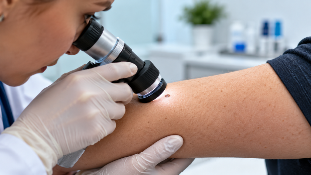
Skin cancer clinics use dermoscopy to examine colours, structures, and patterns beneath the skin’s surface. These details are often difficult or impossible to assess clearly with the unaided eye.
Dermoscopy improves the quality of a clinical examination. It helps a trained clinician decide whether a lesion appears reassuring, needs monitoring, or requires biopsy for laboratory testing.
What Dermoscopy Adds to a Skin Examination
During an examination, dermoscopy may be used alongside the patient’s history, visual inspection, and comparison with other spots. It supports clinical judgment rather than replacing it.
Clinicians may use dermoscopy to:
● Examine structures beneath the skin’s surface
● Assess pigmented and non-pigmented lesions
● Compare one lesion with surrounding spots
● Investigate changes in colour or pattern
● Identify features that deserve closer attention
● Select lesions that may need photography
● Support decisions about monitoring or biopsy
● Reduce avoidable removal of reassuring lesions
Cancer Council Australia states that dermoscopy used by experienced clinicians can increase diagnostic accuracy and reveal features not visible to the naked eye.
Reasons Dermoscopy Is Used in Skin Cancer Clinics
A lesion’s surface appearance provides useful information, but it does not reveal every diagnostic feature. Dermoscopy gives the clinician another layer of visual detail.
The value depends on training and interpretation. The device produces a clearer view, but the clinician must recognise patterns and relate them to the patient’s history and risk.
Magnified Examination
Magnification reveals small structures without cutting the skin, helping clinicians identify reassuring patterns or features that need investigation.
Reduced Surface Reflection
A dermoscope reduces light reflecting from the skin, making deeper colours, vessels, dots, lines, and other structures more clearly visible.
Assessment of Pigmented Lesions
Pigmented spots may contain patterns hidden from normal view. Dermoscopy helps assess symmetry, colour distribution, borders, and internal organisation.
Assessment of Non-Pigmented Lesions
Some skin cancers appear pink, red, pale, or skin-coloured. Dermoscopy may reveal vascular and structural clues beneath the skin on examination.
Comparison With Other Moles
Clinicians compare a lesion with the patient’s other moles. A spot that breaks the usual patternmay deserve closer examination or follow-up.
Support for Monitoring Decisions
Some lesions can be photographed and reviewed later. Dermoscopy for skin lesion assessment at Skin Chx can support careful monitoring decisions over time.
Selection of Lesions for Biopsy
Concerning or uncertain dermoscopic features may lead to tissue sampling. A biopsy allows removed cells to be examined in the laboratory.
| Method | Main purpose | Limitation |
| Visual examination | Reviews the skin surface | Deeper patterns may be unclear |
| Dermoscopy | Magnifies hidden structures | Requires trained interpretation |
| Biopsy | Examines tissue in a laboratory | Involves removing tissue |
Ways Dermoscopy Supports Ongoing Skin Surveillance
Dermoscopy is useful during a single appointment, but it can also contribute to follow-up. Images may allow a clinician to assess whether selected lesions remain stable or develop meaningful changes.
Monitoring is not suitable for every suspicious lesion. A spot with concerning features may need prompt biopsy rather than delayed comparison.
Baseline Documentation
A clear initial image provides a reference for future review. Baseline records improve comparison, particularly when memory or ordinary photographs cannot reliably show small structural details.
Sequential Imaging
Selected lesions may be photographed again after a clinically chosen interval. Side-by-side review can reveal changes in size, colour, structure, or pattern that might otherwise be difficult to detect.
Surveillance for Higher-Risk Patients
People with numerous atypical moles, previous melanoma, or other risk factors may require an individual monitoring plan. Risk influences follow-up frequency, imaging choices, and the threshold for biopsy.
Identification of New Lesions
Comparing current findings with earlier records can help identify spots that were not previously present.
A new lesion deserves contextual assessment, especially when it differs from the patient’s usual mole pattern.
Review of Difficult Areas
The scalp, back, and other hard-to-see areas can be difficult for patients to monitor alone. Clinical examination provides a consistent view of locations that may be missed during self-checks.
Reduction of Unnecessary Excision
Experienced use of dermoscopy may help clinicians recognise reassuring patterns and avoid removing every unusual-looking spot.
Cancer Council reports that dermoscopy has been associated with fewer excisions of benign lesions in dermatologist settings.
Recognition of Its Limits
Dermoscopy cannot guarantee that a lesion is benign or malignant. Uncertain or suspicious findings still require appropriate action, which may include biopsy, referral, treatment, or scheduled review.
What Patients Should Know Before a Dermoscopy Check
Dermoscopy is generally performed during a skin examination and does not involve cutting the skin. The clinician places or holds the device over the lesion and evaluates the magnified view.
Preparation is usually simple, but patients can make the appointment more useful by providing an accurate history.
Before or during the visit:
● Mention lesions that are new or changing.
● Describe bleeding, itching, crusting, or tenderness.
● Share any personal history of skin cancer.
● Mention relevant family history where known.
● Explain previous biopsies or skin treatments.
● Ask which lesions require monitoring.
● Confirm when a follow-up examination is recommended.
● Report further changes rather than waiting silently.
Continue checking your skin between professional appointments if advised. Dermoscopy supports clinical evaluation, but it does not replace awareness of new or changing lesions.
Taking care of your skin also involves building healthy daily habits and staying informed about changes in its appearance. For more practical advice on skincare, grooming, and personal care, explore the latest guides on GroomFade.
Conclusion
Skin cancer clinics use dermoscopy because it reveals colours, structures, and patterns that cannot be assessed as clearly with the naked eye. In trained hands, it supports more accurate decisions about monitoring, imaging, biopsy, or treatment. It does not confirm every diagnosis alone, so clinicians still consider changes, symptoms, risk factors, and pathology when a lesion needs tissue testing.